DNA – You hear it everywhere, but why are scientists so interested in its wonders? Well, DNA is like an encyclopedia. The words inside are composed of only 4 letters:
A (adenine),
G (guanine),
C (cytosine), and
T (thymine).
When scientists examine DNA, they carefully read each and every word inside this complicated encyclopedia. When the words begin to make sense, marvelous things can start to happen.
But how do we retrieve these words or DNA sequences?
Analyzing DNA sequences has aided in several scientific fields since the 1900s. Since then, we have had 3 major DNA sequencing advances, Sanger, FISH, and NGS.
Sanger sequencing
Sanger sequencing was invented by Dr. Frederick Sanger in 1977, a method he would develop for the proceeding 40 years. Also known as the “chain termination method”, it is a method for determining the nucleotide sequence of DNA – the 3 basic steps you can see in the image below:

Though the earliest record of in situ hybridization was founded by Joseph G. Gall and Mary Lou Pardue in 1969, the first fluorescent versions of the technique (FISH) appeared in the 1970s, before direct probe labeling 20 years later.
FISH is a technique that relies on exposing chromosomes to a small DNA sequence called a probe that has a fluorescent molecule attached to it, in order to better detect and locate it. You can see FISH in action below:

NGS was originated through a method known as ‘massively parallel signature sequencing’ (MPSS), which Lynx Therapeutics invented in the 1990s. By 2000, Nick McCooke and his team at Solexa further developed the technology into what you see used in hospitals throughout the world today. NGS is a type of DNA sequencing technology that uses parallel sequencing of multiple small fragments of DNA to determine the sequence. It has 4 major steps (shown below) that allow “high-throughput” via a dramatic increase in the speed (and a decrease in the cost) at which an individual’s genome can be sequenced.

So how do hospitals and clinics best utilize this advance in sequencing technology to improve the standard of care with their patients?
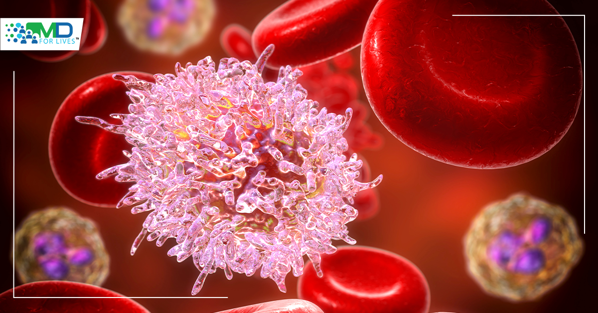
We shall use the example of chronic lymphocytic leukemia (CLL) – a type of cancer in which the bone marrow makes too many lymphocytes (a type of white blood cell). Between 3,800 and 4,500 people are diagnosed with CLL per year in the UK – more than 10 people each day. It affects nearly twice as many men as women, is rare in young people, and is more common in people over 60, with an average age at diagnosis of 72 years.
In the UK, most CLL cases are sequenced using FISH. Generally, a 2-probe set analysis is used, where 2 technologists count 100 nuclei each. The first is a dual-color probe set, consisting of genomic aberrations deletion 11q and deletion 17p. The second is a tri-color probe set that will assess for trisomy 12, 13q14, and 13q34 deletions. Depending on the probe set, the current cut-offs range from 7 to 9%, which correlates to a variant allele frequency of 3 to 55 (VAF is the % of sequence reads observed matching a specific DNA variant divided by the overall coverage at that locus).
The preferred specimen for analysis is peripheral blood (PB). Typically, we perform a direct harvest, but if PB arrives late in the day (>3pm), we would culture it overnight.
In many UK labs, TP53 sequencing is not routinely offered to CLL patients, which means that we are missing a proportion of patients with TP53 mutations that could starkly change their treatment options. Though hospitals can send away larger NGS panels to nearby NGS clinics, this is not always economically available for CLL, due to restricting guidelines and the favoring of these panels for other myeloid malignancies.
There has been a sea change over the last 2 years, however, as more labs have instruments to perform copy number analysis and sequence analysis together, unifying the testing workflow. Labs are now being supplied with automated nucleic acid extraction and liquid handlers for pre-post PCR procedures, as the understanding that public health care systems would benefit from replacing FISH with an assay of similar costs but more range.
In CLL, we are now using more hybrid-capture-based NGS panels. You can see in the image below the copy number variants (CNVs) and single nucleotide variants (SNVs) that are detected.

A December 2021 proof-of-concept analysis performed in the UK, utilizing Oxford Gene technology in 20 patients with CLL, was conducted, where 20 DNA samples previously tested using a different and larger targeted NGS panel and copy number analysis were studied (normal sample data was obtained over 2 NGS runs). Twenty CLL patients who were previously characterized using FISH were then analyzed on an Illumina MiniSeq NGS kit at a high output flow cell. The results can be seen below:

The Fish analysis identified 56$ of nuclei with a heterozygous deletion of TP53, whereas both NGS runs identified the deletion with an estimated tumor content of 57% in each NGS run.
Below you can see a graph indicating the specimens that were a direct preparation (black dots) vs cultured peripheral blood and tumor content by NGS (blue dots). The data shows that there is not a strong bias when the sample is cultured vs directly prepared.

Below, you can see the SNVs identified, with at most 3 mutations identified per patient (with 6 patients with 0 mutations):

In terms of prognosis, you can see the comparison below. After performing the NGS of the 6 patients with a poor prognosis by FISH, 2 were reclassified to intermediate prognosis:

In the future, as NGS becomes more common place, will Hospitals and clinics determine what their in-house limit of detection is? They will perform more NGS on PB specimen that has been fractionated into myeloid and lymphoid fractions. They will run NGS on a non-fractioned specimen and dilute the lymphoid fraction with the myeloid fraction to determine the limit of detection. They will look to lower thresholds for CNV calling without increasing false positive calls and look to verify low-level CNV calls using droplet PCR.
With these steps, hospitals and clinics will be able to increase the number of patients with known copy number variants that could be detected by the NGS platform, which would benefit not only CLL patients but the wider cancer community.


