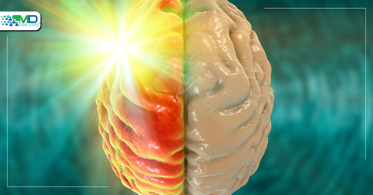
In research on electron transmission to individual DNA molecules, researchers and partners have discovered distinctive fluorescent blinking patterns. These patterns were utilized to locate mRNA glioma point mutations in cell culture. The outcomes of this work may simplify surgical biopsies, enable real-time targeted therapy, and increase our understanding of how cancer develops scientifically. Glioma is a tumor that develops in the brain and spinal cord. Gliomas arise in the gluey supporting cells (glial cells) that surround and support nerve cells. Tumors can be caused by three kinds of glial cells. Gliomas are categorized based on the kind of glial cells involved in the tumor, as well as genetic factors that can help predict how the tumor will behave over time and the therapies that are most likely to succeed.
The surgeon obtains a sample of the problematic tissue during the initial procedure for glioma, a surgical biopsy. The sample is then tested in a lab to determine the kind of cancer (whether benign or malignant) and the type of malignancy. Depending on the outcome of the treatment strategy, you may require a second surgical procedure. However, in a recent work published in Chem, Osaka University researchers and collaborators employed an improved DNA-based fluorescent approach that might help bring real-time cancer detection to medical practice. This discovery provides solutions to long-standing basic scientific concerns and may pave the way for new paths in medical treatment.
Most solid brain tumors, one-third of which are gliomas, are easily detected by standard medical imaging. Unfortunately, two complicated operations are frequently required. However, Japanese researchers may have developed a method to do the initial biopsy, lab testing, and subsequent tumor excision within a single surgical session. Numerous DNA-based biosensors are based on photoinduced electron transfer. The average behavior of numerous molecules, or ensemble measurements, is the foundation for researchers’ knowledge of the kinetics (i.e., rate) of this process. Shuya Fan, the primary author, claims that these metrics conceal the single-molecule behavior that is necessary to comprehend the kinetics of electron transfer. However, this study removes this masking. In order to analyze transitory fluorescence patterns, or “fluorescence blinking”, researchers employed fluorescence correlation spectroscopy. By doing this, they discovered single-molecule chemistry that will enhance diagnostic applications.”
The researchers investigated the correlation between the DNA’s distance and sequence and the kinetics of electron transport in single DNA molecules. Their research was founded on the idea that photoirradiating a fluorescent molecule would trigger electron transfer from the DNA. According to the mathematical method of autocorrelation analysis, the distance between a fluorescent molecule and an electron donor (a hole acceptor) is inversely correlated with the speed of fluorescence blinking.
According to the senior author Kiyohiko Kawai, “unexpectedly, the electron transfer rate for each particular DNA sequence was a unique range of values — a pattern, rather than a specific value.” In cultivated cells, we exploited the corresponding fluorescence blinking to find a glioblastoma point mutation in the mRNA. This research will lead to a better understanding of how point mutations propagate throughout the body. The researchers’ strategy is also appropriate for real-time glioma diagnosis during a surgical biopsy. Therefore, a more practical extension of this study advancement would be focused on cancer therapy without the requirement for several procedures. Possibly as a result of this research, cancer surgery will be less complicated, quicker, and more successful than it is now.

MDForLives is a global healthcare intelligence platform where real-world perspectives are transformed into validated insights. We bring together diverse healthcare experiences to discover, share, and shape the future of healthcare through data-backed understanding.


