Multiple sclerosis (MS) is the most common disabling neurological illness in young adults worldwide. MS may strike at any age, although most patients are diagnosed between the ages of 20 and 50. There are three forms of MS: relapsing, remitting, and progressive, although the course is rarely predictable. Researchers are still trying to figure out what causes MS and why the rate of development is so difficult to predict. The good news is that many persons with MS do not suffer from a significant disability. Most people live a regular or near-normal life. According to research financed by the National MS Society, about 1 million people in the United States have MS. Globally, the expected number of persons living with MS has risen to 2.8 million by 2020. The estimate is 30% higher than in 2013.
Damage to the myelin in the central nervous system (CNS) — and to the nerve fibres themselves — interferes with nerve signal transmission between the brain and spinal cord and other regions of the body in multiple sclerosis. MS symptoms cannot be predictable and vary from person to person. Each person’s symptoms might alter or fluctuate over time, and no two people have precisely the same symptoms. Exercise has been proven in studies of patients with multiple sclerosis to aid with fatigue and depression, enhance strength, and boost involvement in social activities.
Multiple sclerosis (MS) causes a wide range of symptoms that have a significant negative impact on patients’ well-being and quality of life.

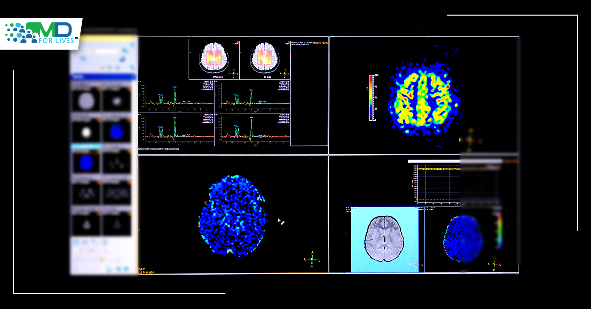
MS does not have any specialised testing. Instead, a differential diagnosis, or ruling out other illnesses that may generate similar signs and symptoms, is frequently used to confirm a diagnosis of multiple sclerosis. Blood tests, spinal taps (lumbar punctures), MRIs, and evoked potential testing are some of the most common tests performed. The diagnosis of relapsing-remitting MS is usually simple, based on a pattern of symptoms that are consistent with the condition and confirmed by brain imaging studies such as MRI. Multiple sclerosis (MS) should be detected and treated as soon as possible to prevent the illness from progressing. Magnetic resonance imaging (MRI) is an important tool in this procedure. A novel MRI approach was employed at MedUni Vienna as part of a research effort that might lead to a faster evaluation of disease activity in MS. The work was recently published in the journal Radiology by a research team led by Wolfgang Bogner from MedUni Vienna’s Department of Biomedical Imaging and Image-guided Therapy.
Although standard MRI can identify abnormalities in the brain, scientists are working on ways to detect alterations at a microscopic or biochemical level. Proton MR spectroscopy has been recognised as a potentially useful instrument for this aim. In their recently published work, the research team used this method to go one step further. The researchers compared the neurochemical alterations in the brains of 65 MS patients with those of 20 healthy controls using MR spectroscopy with a 7-tesla magnet. Since its commissioning in 2008, MedUni Vienna’s Center of Excellence for High-Field MR has been utilised for scientific investigations, such as brain studies, at MedUni Vienna’s Center of Excellence for High-Field MR.
By detecting and anticipating changes, researchers from MedUni Vienna have identified MS-relevant neurochemicals using 7-tesla MRI. It allowed them to see abnormalities in the brain in areas that seemed normal on traditional MRI imaging. These discoveries might have a big impact on how MS sufferers are treated in the future. Some of the neurochemical changes they were able to see with the new approach occur early in the course of the disease and may not only correlate with impairment but also predict disease progression.
There will be more clinical trials and advances in the future. Before these findings may be used in therapeutic settings, more study is required. The findings suggest that 7-tesla spectroscopic MR imaging might be a useful new tool in the diagnosis and treatment of multiple sclerosis patients. If the findings are validated in other research, this novel neuroimaging approach might become a common imaging tool for MS patients’ initial diagnosis, monitoring disease activity, and therapy. The approach is now only accessible for research reasons on Austria’s sole 7-Tesla MRI scanner at MedUni Vienna. The researchers are currently striving to improve the novel technology so that it may be used in normal clinical MRI scanners.
Reference:
- https://pubs.rsna.org/doi/full/10.1148/radiol.210614
- https://www.ncbi.nlm.nih.gov/pmc/articles/PMC7720355/#:~:text=The%20estimated%20number%20of%20people,%2C%2035.95%5D%20per%20100%2C000%20people.
- https://www.nationalmssociety.org/What-is-MS/Who-Gets-MS
- https://www.mayoclinic.org/diseases-conditions/multiple-sclerosis/diagnosis-treatment/drc-20350274


