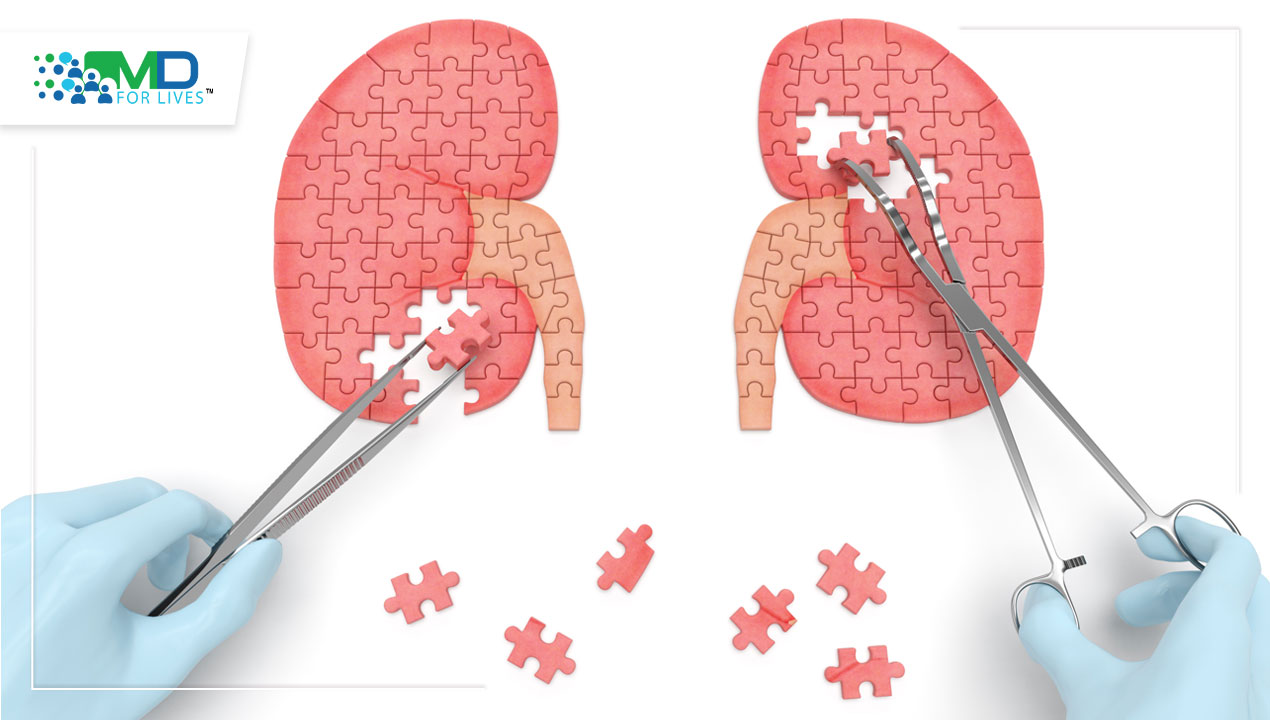
To put it simply, renal diagnostics holds the key to a future where patients with renal diseases find hope in every test result. It is a combination of procedures that helps clinicians obtain valuable insights into renal function and anatomy, enabling early detection and targeted treatment of renal diseases. In essence, renal diagnostics promises better management, improved quality of life, and brighter tomorrows for those battling renal conditions.
However, cutting-edge renal diagnostics is not really possible without advanced imaging.
Offering detailed visualization of renal anatomy, structural abnormalities, and functional integrity, renal imaging plays a pivotal role in guiding targeted therapeutic interventions. Today, various imaging modalities are utilized depending on the clinical scenario, each offering unique advantages.
Through this blog, let us delve into all the current modalities and upcoming innovations that are setting new milestones in the accuracy of renal imaging.
Advancements in Imaging Techniques
Despite the availability of widely used imaging modalities, their limitations have prompted researchers to explore new innovations.
For instance,ultrasound, while advantageous for its non-invasive nature and real-time imaging, is limited by operator-dependence, reduced sensitivity for small masses, and limited anatomical detail compared to CT or MRI.
Even Computed Tomography (CT) scans, which facilitate accurate characterization and localization of lesions within the kidneys, involve exposure to ionizing radiation and carry potential health risks for patients.
Likewise, nuclear medicine techniques, including renal scintigraphy, diuresis renography, and positron emission tomography (PET) scans, offer valuable insights into kidney structure and function but pose the risk of radiation exposure and the need for contrast agents.
To overcome the shortcomings of these existing imaging techniques, clinicians are increasingly adopting new imaging modalities that are far more technologically advanced and risk-free than their predecessors.
Contrast-Enhanced Ultrasound (CEUS)
Contrast-enhanced ultrasound (CEUS) has emerged as a cost-effective and efficient imaging method for characterizing indeterminate renal lesions, offering excellent visualization of the renal vasculature and accurate assessment of focal infarction and cortical necrosis. CEUS is relatively easy to learn and provides a simple decision-making process. It is a valuable tool that enhances higher diagnostic accuracy in renal lesion assessment than CT and MRI.
CFE-MRI
Cationic ferritin-enhanced magnetic resonance imaging (CFE-MRI) represents another advancement in imaging techniques for the kidneys. By utilizing cationic ferritin, which binds to anionic proteoglycans in the glomerular basement membrane and surface glycocalyx of the glomerular endothelium, CFE-MRI enables the visualization and assessment of glomerular number, function and pathology with enhanced sensitivity and specificity. This imaging technique offers a non-invasive and innovative approach to evaluating renal structure and function at the microstructural level.
3D Imaging
In recent years, patient-specific 3D models have become indispensable for preoperative planning, particularly for individuals with renal tumors who may benefit from minimally invasive or nephron-sparing procedures. These models aim to improve surgical accuracy and patient safety while minimizing intraoperative complications and reducing the duration of surgery. These models come in various forms including virtual, printed, or augmented reality (AR), each offering unique advantages in visualizing anatomical structures and facilitating surgical decision-making. Virtual models are created using 3D modeling tools and computer graphics techniques, while printed models are fabricated via 3D printing from digital files, providing tangible representations for surgeons to handle. Augmented reality (AR) combines real-time imaging with virtual overlays, allowing surgeons to augment anatomical structures with additional information, aligning preoperative images with the surgical field for enhanced intraoperative guidance.
Ushering in New Breakthroughs in Renal Imaging
Regularizing the Use of AI and ML
Artificial Intelligence (AI) and Machine Learning (ML) are poised to enhance renal imaging by automating image interpretation, improving diagnostic accuracy, and predicting disease progression and outcomes. Analyzing vast amounts of imaging data alongside clinical parameters, these technologies can help doctors devise personalized treatment plans and stratify risk levels for patients with renal diseases. Integrating AI-driven solutions with electronic health records can further streamline workflows and facilitate data-driven decision-making in renal care.
Molecular Imaging
Molecular imaging holds significant potential for targeted diagnosis and therapy in renal pathophysiology by allowing visualization of specific molecular processes within the kidney. Recent advancements in imaging agents and techniques enable the detection of molecular biomarkers associated with various renal diseases, facilitating early diagnosis and personalized treatment strategies. By leveraging molecular imaging, clinicians can gain insights into the underlying molecular mechanisms of renal conditions and optimize therapeutic interventions for improved patient outcomes.
Portable and Wearable Imaging Technologies
The growing adoption of portable ultrasound and other wearable imaging modalities has revolutionized remote monitoring in renal diagnostics by enabling real-time imaging beyond traditional clinical settings. These technologies offer convenient and non-invasive means to assess renal function, detect abnormalities, and monitor disease progression in remote or resource-limited areas. Their portability enhances accessibility to renal imaging, facilitating early intervention and personalized management of renal conditions, ultimately improving patient outcomes.

For physicians looking to stay updated with the latest advancements in renal imaging techniques, enhancing their diagnostic capabilities and patient care, MDForLives, the largest medical survey platform, offers invaluable resources and support in the field of imaging in renal diagnostics. MDForLives provides comprehensive insights into current modalities and future directions through informative articles, webinars, and case studies.
Additionally, MDForLives offers paid surveys for healthcare professionals, allowing them to share their expertise on healthcare topics while being compensated for their time and insights. Moreover, physicians can further expand their professional reach by writing blogs and case studies and receiving compensation for their contributions. Finally, MDForLives offers physicians the chance to participate as paid speakers or participants in webinars, fostering knowledge exchange and professional development within the medical community.
Join MDForLives now and be at the forefront of this transformative period in the medical industry.


