

What if researchers could switch a specific group of brain cells on or off and see what happens next? That once sounded like science fiction. Today it describes optogenetics, a technology that combines genetic targeting with light-sensitive proteins to control selected cells with precision.
In October 2026, Karl Deisseroth, Peter Hegemann, and Georg Nagel received the Nobel Prize in Physiology or Medicine for discoveries involving light-gated ion channels and optogenetics.
The importance goes beyond making neurons respond to light. Optogenetics lets researchers move from observing brain activity to testing cause and effect, asking whether a specific group of neurons or circuit actually contributes to movement, memory, behavior or disease. That shift has changed brain mapping and how scientists study disorders including Parkinson’s disease and epilepsy.
But what is optogenetics, how does it work, and how close is it to becoming part of neurological disease treatment?
Optogenetics began with a light-sensitive protein
The story starts far from the human brain. In the early 2000s, researchers studying the green alga Chlamydomonas reinhardtii identified a family of light-responsive proteins called channelrhodopsins. One of them, channelrhodopsin-2, proved especially important: when light hits it, it rapidly opens an ion channel, letting charged particles flow across the membrane and change the cell’s electrical state.
Neurons communicate electrically, which raised a question: could a light-sensitive protein be placed into selected neurons to control them? In 2005, researchers showed that neurons expressing channelrhodopsin-2 could be made to fire in response to precisely timed light pulses, within milliseconds, matching the timescale of neural signals. That is when the foundation of modern optogenetics was established.
How does optogenetics work?
Optogenetics combines two components; genetic targeting and light. Selected cells are made to produce a light-sensitive protein called an opsin, and different opsins respond to specific wavelengths; when researchers deliver light, illumination can increase or suppress electrical activity depending on the opsin and design. In a simpler way, it is like installing a light-responsive switch in selected cells.
The advantage is precision: researchers need not stimulate every neuron in a region but can target defined cell types or connections and change their activity at controlled moments. This is especially useful when neighboring neurons perform very different functions.
Brain mapping moved from correlation to cause
Traditional brain mapping can show that a region becomes active during movement, memory, fear or another function, but activity alone does not prove the region caused the response. Optogenetics lets scientists test that relationship directly: they can activate one population of neurons and observe whether behavior changes or inhibit another and assess which function is affected. Brain mapping can therefore move from “Where is activity happening?” to “Which cells and circuits actually contribute to this function?”
Memory research offers a striking example. In 2012, scientists identified hippocampal neurons active while mice formed a fear memory, then reactivated those cells with light, producing behavior associated with recall. The study did not create a treatment; its significance was methodological, showing that a specific population of neurons formed part of the circuitry supporting a memory.
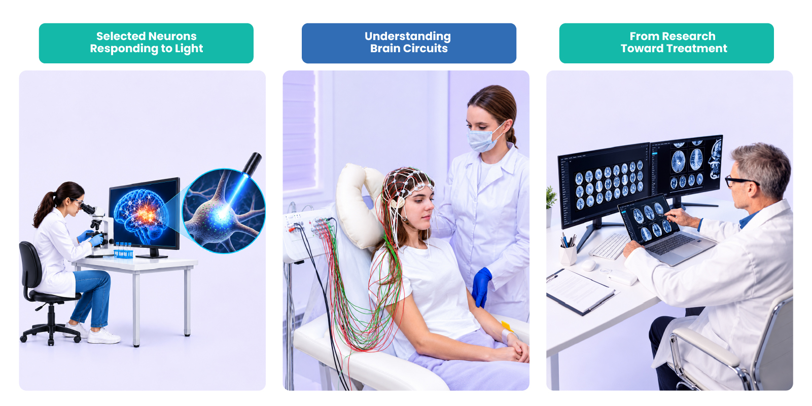
Brain circuits can reveal disease mechanisms
The same approach has helped researchers investigate neurological and psychiatric disorders. In Parkinson’s disease, movement depends on interconnected pathways within the basal ganglia. A 2010 mouse study used optogenetics to activate different pathways selectively: one produced parkinsonian-like movement abnormalities, while another improved movement and reversed several motor deficits in the model. This does not make optogenetic therapy an established Parkinson’s treatment, but it showed that different circuits have different causal roles in movement.
Similar research has explored circuits involved in memory, reward, sleep, and depression-related behaviour. That knowledge can matter clinically even if light never treats the patient: a circuit linked to a symptom may become a target for another approach, such as deep-brain stimulation or medicines.
Epilepsy shows why timing also matters
Epilepsy illustrates another advantage: control over both where and when neural activity is modified. A seizure involves abnormal electrical activity, but broadly suppressing brain activity can interfere with normal function. In a 2013 mouse model of temporal-lobe epilepsy, researchers built a closed-loop system that detected spontaneous seizures and delivered optogenetic stimulation to selected hippocampal cells, stopping seizures in the model. This showed that abnormal brain activity could potentially be detected and modified in real time. It is still far from an established human epilepsy treatment, but it shows why precise circuit control matters when abnormal activity appears unpredictably, and timing matters as much as location.
Optogenetic therapy has reached humans
Moving from research to human treatment is much harder. Human cells must safely acquire a light-sensitive protein, and researchers must deliver enough light to activate them without unacceptable harm. The retina is a useful starting point because it is naturally accessible to light.
Optogenetic therapy depends on delivering genetic instructions that can make selected cells responsive to light. Explore gene therapy, its uses, mechanisms, and what to expect.
In 2021, researchers reported partial recovery of visual function in a patient with advanced retinitis pigmentosa, after gene delivery made retinal ganglion cells light-sensitive and specialized goggles supplied the stimulation. The evidence has since moved beyond one case. A 2026 New England Journal of Medicine open-label study evaluated optogenetic therapy in 10 people blinded by advanced retinitis pigmentosa. Seven showed increased light sensitivity, and six reached the study’s definition of clinically meaningful improvement. Safety was the primary outcome: most ocular adverse events were mild or moderate, and one severe transient event after the injection resolved within minutes. These findings are an important step, though not a sign that optogenetics is ready for routine treatment of disorders deep within the brain.
The brain remains the harder target
Applying optogenetics inside the human brain adds challenges. Researchers must identify the correct cells, deliver genetic material safely, and get light to the intended region, while gene expression, immune responses, light-delivery devices and regulation all need consideration. Neurological disorders also rarely involve one isolated switch; conditions like Parkinson’s disease, epilepsy and depression involve networks of interacting circuits, and a circuit contributing to a symptom may also serve normal functions. Precision therefore means more than location; which cells are targeted, where, and when.
Precision is becoming increasingly important as researchers move toward treatments that target specific cells, pathways, and disease mechanisms. Explore how precision medicine can improve treatment and care.
A 2025 translational roadmap described two routes from optogenetics to clinical care. One is direct translation, using the technology itself as treatment, and the other one is indirect translation, using its discoveries to identify cells or circuits that other treatments, such as electrical stimulation or medicines, can target. For many brain disorders, the indirect route may shape practice long before direct therapy becomes feasible.
Closing perspective: illuminating cause, not just activity
Optogenetics is often described as controlling the brain with light. The deeper truth is that a light-sensitive protein discovered in algae became a tool able to control selected neurons within milliseconds. It helped move brain mapping from observation toward causation, revealing how specific circuits contribute to movement, memory, behavior, and disease, and has begun to enter human research through experimental retinal therapy.
The future is therefore likely to follow two paths: one direct, determining whether optogenetic therapy can become safe and practical for selected human conditions, and one perhaps equally important, using optogenetics research to identify the precise brain circuits that other treatments should target. The Nobel Prize recognizes how dramatically optogenetics has changed neuroscience; its eventual clinical legacy may depend on how well that understanding translates into safer, more precise treatment.