Kidney stone disease (also called renal calculi, nephrolithiasis, or urolithiasis) is a common urological condition occurring due to the deposition of minerals and salts in the kidneys. It affects approximately 0.9% of the United States population, with a steady increase in incidence within the past four decades.
Surgical procedures are often required to remove primary stones in the kidneys or ureter that are symptomatic and larger than 6mm. Meanwhile, secondary stones that are small and asymptomatic are typically left behind. Ideally, these stones can pass through the ureter without causing any complications. Current guidelines only suggest active monitoring for the management of secondary kidney stones.
However, studies have shown that patients with untreated asymptomatic stones and fragments can have approximately a 50% chance of relapse within 5 years post-surgery. The decision to remove the stones is left in the hands of patients and their urologists.
A recent study published in the New England Journal of Medicine considered the problem of what to do with small, asymptomatic kidney stones that are found during the detection of primary kidney stones.

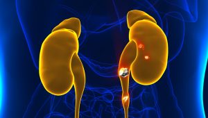
Caption: Primary kidney stones can co-occur with smaller secondary stones that can form in the ipsilateral or contralateral kidneys.
Symptomatic vs Asymptomatic Kidney Stones
Kidney stone formation is influenced by various risk factors such as diet, obesity, intake of certain supplements and medications, and certain medical conditions.
Kidney stones are usually asymptomatic until they move within the kidney or pass into the ureters that connect the kidneys to the bladder. In this case, patients can experience severe and sharp pain radiating from the lower abdomen and groin region and a burning sensation while urinating. Symptomatic kidney stones can also cause urinary tract infections and impairment of kidney function if not treated.
On the other hand, asymptomatic kidney stones are generally smaller (raging less than <6mm) and may not be associated with the risk of developing clinical complications. Preoperative imaging using computed tomography (CT) is generally used to identify the location and stone burden in patients and inform treatment decisions.
Treatments for Kidney Stones
U.S. and European guidelines suggest ureteroscopy (URS), percutaneous nephrolithotomy (PNL), or shock wave lithotripsy (SWL) for the surgical removal of symptomatic kidney stones. The choice of treatment depends on the location, size, and composition of the kidney stones as well as the burden of symptoms and presence of any patient comorbities. There is no consensus regarding the treatment of secondary kidney stones except for active monitoring.
Clinical trial on the removal of Asymptomatic Kidney Stones
A recent multicenter, randomized, controlled trial investigated outcomes from removing secondary stones in patients who were undergoing endoscopic removal of larger primary kidney stones. In the study, 38 patients had both their primary and secondary stones removed (treatment group) while 35 patients had only their primary stones removed (control group). Primary stones were defined as stones in the kidney or ureter that were symptomatic or that were judged as presenting a high risk of adverse clinical events. Secondary stones were smaller or equal to 6mm in diameter and located in the contralateral kidney or in either kidney if the primary stone was in the ureters.
The risk of relapse was the study’s primary outcome. Patients were considered to have relapsed if they either had an emergency department visit due to new stone(s) forming at the location of the initial asymptomatic stone, or follow-up surgery to remove stones on the trial side, or growth of the initial secondary stone.

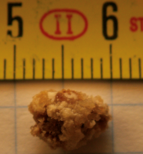
Caption: A kidney stone.
During follow-up (mean of 4.2 years), the treated patients had an 82% lower risk of relapse (hazard ratio, 0.18; 95% confidence interval, 0.07 to 0.44). The mean time to relapse in the treatment group was 75% longer than in the control group; when stone growth was excluded from the primary outcome, treated patients had a 36% longer time to relapse compared with control patients.
Other Study Outcomes:
The authors also considered immediate and near-term effects of the additional surgical treatment. They found that five patients in the treatment group (13%) and four patients (11%) in the control group had an emergency department visit within 2 weeks following surgery; all these visits were due to stent pain. Overall, the authors reported an average increase of 25.6 minutes of surgery time with the removal of secondary kidney stones in the treatment group. The cost of the additional surgery time will likely be much smaller than the cost of emergency department visits and subsequent surgeries.
The authors state that these findings may justify the need for prophylactic surgery for secondary stones in all patients undergoing endoscopic removal of kidney stones. Limitations in this study include the relatively small pool of patients, which included few individuals from non-white groups and no Hispanic patients.
The clinical trial was funded by the National Institute of Diabetes and Digestive and Kidney Diseases and the Veterans Affairs Puget Sound Health Care Systems.

MDForLives is a global healthcare intelligence platform where real-world perspectives are transformed into validated insights. We bring together diverse healthcare experiences to discover, share, and shape the future of healthcare through data-backed understanding.


