

PSMA PET scan for prostate cancer is transforming how clinicians detect, stage, and monitor disease progression. Traditional imaging methods such as CT scans and bone scans often struggle to identify small or early metastatic lesions, particularly at low PSA levels. PSMA PET imaging addresses this gap by targeting prostate-specific membrane antigen, a protein highly expressed on many prostate cancer cells.
Over the past few years, landmark studies including the ProPSMA, CONDOR, and OSPREY trials have demonstrated significantly improved detection accuracy compared to conventional imaging approaches. As precision oncology advances, PSMA PET CT scan technology is becoming increasingly important in treatment planning, recurrence monitoring, and personalized prostate cancer management.
What is Prostate-Specific Membrane Antigen PET? (PSMA PET Scan)
A PSMA PET scan is a molecular imaging technique used primarily in prostate cancer diagnosis and staging. The scan combines positron emission tomography with a radioactive tracer designed to bind to prostate-specific membrane antigen.
The PSMA PET scan full form is:
Prostate-Specific Membrane Antigen Positron Emission Tomography
When PET imaging is combined with CT, the procedure is called a PSMA PET CT scan.
Unlike standard PET imaging that commonly evaluates glucose metabolism, PSMA PET imaging directly targets proteins present on prostate cancer cells. This improves specificity and increases the ability to identify cancer spread at earlier stages.
Commonly Used PSMA Tracers
| Tracer | Type | Regulatory Milestones |
| Gallium-68 PSMA-11 | Ga-68 tracer | FDA approved in 2020 |
| Pylarify | F-18 tracer | FDA approved in 2021 |
| Posluma | F-18 tracer | FDA approved in 2023 |
These tracers help improve visualization of:
- Lymph node metastases
- Bone metastases
- Biochemical recurrence
- Small metastatic lesions

How does PSMA PET Work?
The scan works by injecting a small amount of radioactive tracer into the bloodstream. The tracer selectively binds to prostate cancer cells expressing PSMA.
Once attached, the tracer emits signals that are detected by PET scanners, producing highly detailed molecular images.
The Process Involves:
- Intravenous tracer injection
- Waiting period for tracer uptake
- PET CT imaging acquisition
- Specialist interpretation of results
This molecular targeting approach allows clinicians to identify disease that may not yet be visible through structural imaging.
What PSMA PET Imaging Can Detect
- Small lymph node involvement
- Bone lesions
- Recurrent disease at low PSA levels
- Treatment response patterns
- Oligometastatic disease
PSMA Biology: Why the Scan Matters
PSMA is a cell-surface protein highly overexpressed in many prostate cancer cells.
Importantly:
- Expression often increases in aggressive disease
- Higher PSMA expression may correlate with metastatic progression
- The protein is minimally expressed in most normal tissues
This makes PSMA an effective molecular target for imaging.
The same targeting mechanism is also being explored in PSMA-based radioligand therapies such as Lutetium-177 PSMA therapy.
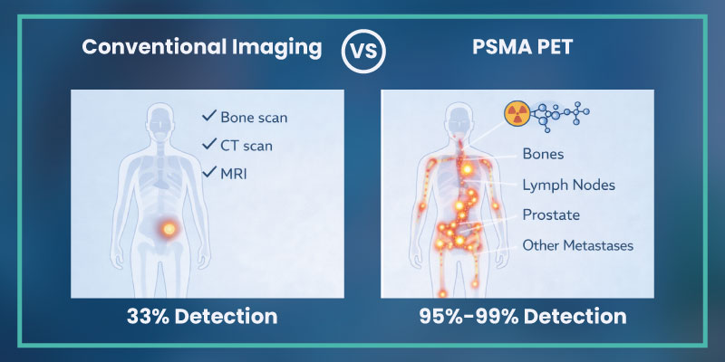
How Is PSMA PET Imaging Different from Current Prostate Cancer Imaging?
Traditional imaging methods primarily evaluate structural abnormalities.
Conventional Imaging Limitations
| Imaging Type | Primary Limitation |
| CT Scan | Limited sensitivity for small metastases |
| Bone Scan | Lower specificity |
| MRI | Primarily anatomical evaluation |
| Conventional PET | Less prostate-specific targeting |
PSMA PET Imaging Advantages
| Feature | PSMA PET Scan |
| Molecular targeting | Yes |
| Detects low-volume disease | Higher sensitivity |
| Detects recurrence at low PSA | Yes |
| Micro-metastatic detection | Improved |
| Treatment planning support | Strong |
The ProPSMA Trial
The ProPSMA trial, published in 2020, demonstrated that PSMA PET imaging showed significantly greater accuracy than conventional imaging in high-risk prostate cancer staging.
Key findings included:
- Improved detection accuracy
- Better identification of metastatic disease
- Fewer equivocal findings
- Greater impact on treatment decisions
This study played an important role in expanding clinical adoption globally.
Duration: How Long Does a PSMA PET Scan Take?
Patients commonly ask: how long does a PSMA PET scan take?
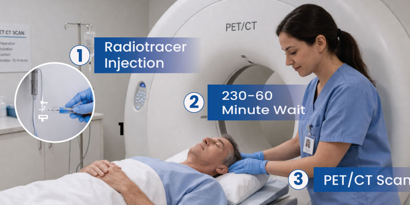
The complete appointment generally lasts between 2 and 3 hours.
Typical Timeline
| Step | Estimated Time |
| Registration and preparation | 15 to 30 minutes |
| Tracer injection | 5 minutes |
| Uptake waiting period | 45 to 60 minutes |
| Imaging scan | 20 to 40 minutes |
| Total duration | Approximately 2 to 3 hours |
The imaging portion itself is usually under 30 minutes.
Procedure: How PSMA PET Scan Works
The PSMA PET scan procedure follows a standardized workflow.
Step-by-Step Procedure,
1. Pre-Scan Assessment: Patients review medical history and preparation instructions.
2. Radiotracer Injection: A small amount of tracer is injected intravenously.
3. Uptake Period: Patients wait while the tracer circulates and binds to PSMA-expressing cells.
4. PET CT Imaging: The patient lies on a scanning table while whole-body imaging is performed.
5. Image Interpretation: Nuclear medicine specialists analyze the scans and generate a report.The procedure is non-invasive and generally well tolerated.
PSMA PET Scan Preparation
PSMA PET scan preparation is relatively straightforward.
Preparation Guidelines
- Stay hydrated before the scan
- Avoid strenuous exercise before imaging
- Follow medication instructions from the imaging center
- Inform clinicians about allergies or medical conditions
Fasting is not always required, although protocols may vary by facility.
What Happens During the PSMA PET Scan?
During the scan, patients lie still on a motorized table that moves through the scanner.
The PSMA PET scan machine detects signals emitted by tracer-bound cells and converts them into detailed images.
Patients Can Expect:
- Minimal discomfort
- No surgical procedures
- Quiet imaging environment
- Continuous monitoring by imaging staff
Movement should be minimized to ensure image clarity.
Clinical Benefits of PSMA PET Imaging
PSMA PET imaging has significantly influenced prostate cancer management strategies.
Key Clinical Benefits ,
- More Accurate Staging: Clinicians can identify metastatic disease earlier and more precisely.
- Better Detection of Biochemical Recurrence: PSMA PET scan for prostate cancer recurrence can detect lesions even at very low PSA levels.
- Improved Radiation Planning: Targeted treatment planning becomes more accurate.
- Reduced Unnecessary Procedures: Improved staging may help avoid unnecessary surgery or broader radiation exposure.
- Support for Personalized Treatment Decisions Findings influence: Surgery decisions ,Salvage therapy planning ,Systemic therapy selection ,Metastasis-directed treatment
Accuracy of PSMA PET Scan
Multiple studies have shown superior sensitivity and specificity compared with conventional imaging.
Detection Performance: Detection rates increase as PSA levels rise, but PSMA PET imaging remains valuable even at low PSA levels.
Reported Clinical Impact: Studies have shown:
- Changes in treatment planning in 25 to 40 percent of cases
- Improved identification of oligometastatic disease
- Upstaging of apparently localized disease
Important Trials Supporting Accuracy
| Trial | Key Contribution |
| ProPSMA | Improved staging accuracy |
| CONDOR | Recurrence detection at low PSA |
| OSPREY | Improved localization and staging |
Role of PSMA PET in Biochemical Recurrence
Biochemical recurrence occurs when PSA levels rise after surgery or radiation therapy.
One of the major strengths of PSMA PET imaging is the ability to localize recurrent disease even when PSA levels are low.
Why This Matters
- Earlier intervention opportunities
- Better salvage therapy planning
- Reduced uncertainty after rising PSA
This capability has made PSMA PET increasingly valuable in recurrent prostate cancer management.
Who Should Consider PSMA PET?
PSMA PET is commonly considered for:
- High-risk newly diagnosed prostate cancer
- Biochemical recurrence after treatment
- Rising PSA with negative conventional imaging
- Pre-salvage therapy evaluation
- Assessment of metastatic spread
Eligibility depends on clinical assessment, regional availability, and regulatory approvals.
PSMA PET Scan Results
Scan results are interpreted by nuclear medicine physicians and radiologists.
Reports Typically Include:
- Location of lesions
- Number of suspicious areas
- SUV values indicating tracer uptake intensity
- Evidence of metastasis or recurrence
Understanding SUVmax
SUVmax refers to standardized uptake value maximum.
Higher SUV values may indicate:
- Increased tracer uptake
- Greater PSMA expression
- Higher suspicion for active disease
However, SUV interpretation must always be correlated with clinical context.
PSMA PET Scan Side Effects
Patients frequently ask about side effects of procedures.
The scan is generally considered safe and well tolerated.
Possible Side Effects
- Mild discomfort at injection site
- Rare allergic reactions
- Minimal radiation exposure
Serious complications are uncommon.
Limitations of PSMA PET Imaging
Although highly effective, PSMA PET imaging has limitations.
Important Considerations
- Not all prostate cancers strongly express PSMA
- False positives can occasionally occur
- Availability varies by region
- Cost remains a barrier in some healthcare systems
Interpretation should always occur within multidisciplinary clinical assessment.
PSMA PET Scan Cost and Availability by Country
PSMA PET scan cost continues to rise globally.
United States
- Approximate cost: $3,000 to $7,000
- Medicare coverage depends on indication
- Insurance coverage varies
Patients often ask:
- Is PSMA PET scan covered by Medicare?
- What is the PSMA PET CT scan cost?
Coverage policies continue to evolve.
India
- Approximate range: ₹25,000 to ₹60,000
- Available in major oncology centers
- Regulated under AERB guidelines
United Kingdom
- NHS availability varies regionally
- Private scan costs may range from £1,500 to £3,000
- NICE guidance continues to evolve
Germany
- Available in major university hospitals
- Coverage may vary between statutory and private insurance systems
France
- Access varies across public and private systems
- Reimbursement policies continue to expand
Italy
- Availability differs by region
- Increasing integration into advanced oncology centers
Does PSMA PET Scan Detect Other Cancers?
Although PSMA imaging is primarily used in prostate cancer, PSMA expression has been observed in:
- Certain renal tumors
- Some salivary gland cancers
- Selected other malignancies
However, the strongest validated clinical role remains in prostate cancer imaging.
Treatment After PSMA PET Scan
Treatment depends on imaging findings.
Potential Next Steps
| Imaging Finding | Potential Treatment Direction |
| Localized disease | Surgery or radiation |
| Oligometastatic disease | Targeted metastasis therapy |
| Widespread metastasis | Systemic therapy |
| Biochemical recurrence | Salvage therapy |
PSMA PET functions as a decision-support tool in multidisciplinary oncology care.
Read about Brachytherapy for Prostate Cancer
The Future of PSMA PET-CT: How PSMA Revolutionizes Staging for Prostate Cancer
PSMA PET imaging is increasingly central to precision oncology.
Emerging Developments ,
- AI-Assisted Imaging Analysis:Artificial intelligence tools may improve lesion detection and interpretation consistency.
- PSMA Theranostics: PSMA-targeted radioligand therapy, including Lutetium-177 PSMA treatment, is rapidly expanding.
- Earlier Detection Strategies: Future studies are exploring lower PSA thresholds and earlier intervention.
- Personalized Oncology Planning: Molecular imaging may further support individualized treatment mapping. The future direction combines imaging, targeted therapy for cancer, and data-driven oncology.
Conclusion
PSMA PET scan technology represents one of the most important advances in modern prostate cancer imaging. By combining molecular targeting with high-resolution imaging, it improves staging accuracy, recurrence detection, and treatment planning.
As clinical evidence continues to expand through studies such as ProPSMA, CONDOR, and OSPREY, PSMA PET CT scan applications are becoming increasingly integrated into routine prostate cancer care pathways.
The convergence of molecular imaging, precision oncology, and targeted therapy is redefining how clinicians approach diagnosis and long-term prostate cancer management.